Probably one of the most common questions I am asked by patients is “do I have any blockages in my heart doctor?”
I usually encounter this question after patients have undergone certain investigations. It is an important question no doubt, as more and more people are now concerned about heart disease.
I thought I would briefly discuss what a ‘heart blockage’ is, and how we detect it through certain tests. I will also discuss the best ways to detect heart blockages as well.
What Is A Heart Blockage?
A heart blockage is a term that is used by patients when they wish to know if they are at risk of having a heart attack. What I have found over the years is that when patients undergo cardiac tests, the one thing they almost always ask me is whether they have blockages in the heart.
I often presume that this means whether the heart arteries are blocked or not.
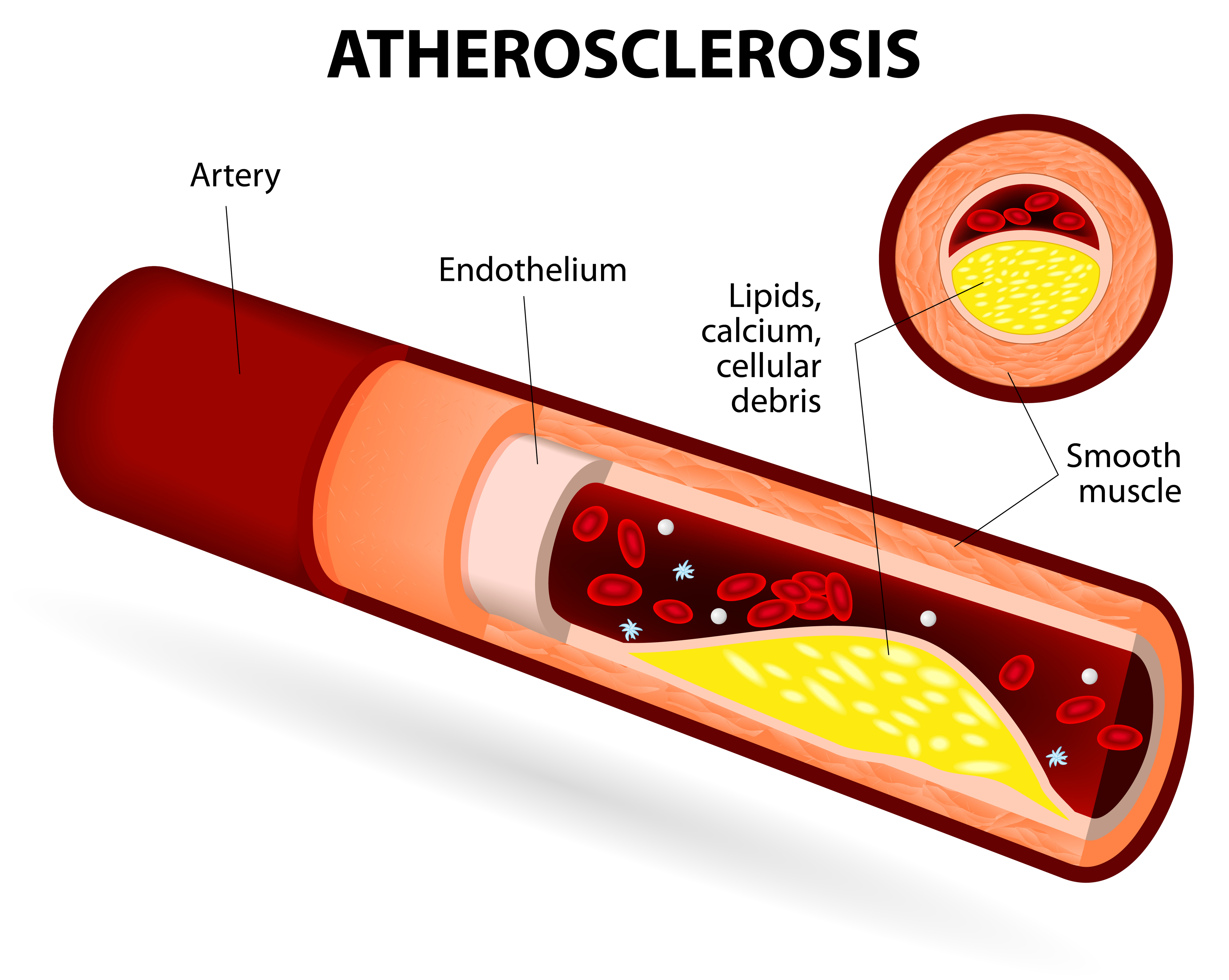
Narrowing of the heart arteries is usually confused with blocked arteries. When arteries are narrowed, the quantity of blood flowing to the heart muscle in times of need is reduced. Narrowing of the arteries is due to a process called atherosclerosis.

For example, the heart requires more blood during exercise. The extra blood gives it the nourishment it needs to continue functioning efficiently.
However, if there are narrowing of the blood vessels that supply these nutrients, the heart begins to struggle.
A ‘narrowing’ and ‘blockage’ describe different conditions. When doctors describe blockages, we mean the heart artery is 100% blocked. A narrowed artery can only be 80% narrowed, but not blocked.
Detecting these ‘narrowings’ or ‘blockages’ can be achieved through certain investigations. Here, I will discuss a step wise approach cardiologists and physicians follow when detecting ‘heart blockages’ for the first time.
For the purpose of this article and for better understanding, I will use the term heart blockages to describe narrowing of the blood vessels.
How Are Heart Blockages Detected?
The first way to detect whether a heart artery is blocked or not is by symptoms. Patients who have severely narrowed arteries can experience a number of symptoms. I have listed these in the table below.
1. Chest pain affecting the upper part of the chest accompanied with sweating and nausea
2. Radiation of pain to the jaw or left arm
3. Reduced ability to exercise due to pain or breathless
4. Breathlessness worsening as time passes
5. Palpitations accompanying the chest pain
However, some individuals may not experience any symptoms whatsoever. This is particularly common in people who have diabetes.
That being said, sometimes people tend to ignore some of the symptoms I have mentioned above. Many feel their chest pain is due to ‘gas’.
If you ever have chest pain of any sort and are even the slightest bit concerned about it, make sure you see your doctor.
Electrocardiogram
An electrocardiogram, or an ECG, is a good way of determining if there has been any damage to the heart muscle due to heart blockages. It is sometimes called a heart tracing.
Doctors will look for specific changes in your heart tracing that would indicate the presence of a damaged heart muscle.

However, the ECG can sometimes be normal. This is not uncommon.
When the ECG is normal, it comes down to symptoms and clinical suspicion. If your doctor feels that your symptoms are due to reduced blood flow to your heart, you may have additional investigations arranged.
Treadmill Testing
I have discussed treadmill tests in detail here.
While a treadmill test is a good way of screening patients for heart disease and blockages, it does not give 100% accurate results.
We conduct treadmill tests in our clinic all the time, and on rare occasions the test can be negative despite the patient having heart disease.
This is what a patient walking on a treadmill looks like –
Clinical studies have shown that 1 out of 5 cases of heart disease can be missed with a treadmill test. In other words, 20% of patients will have a normal study even if their arteries are narrowed.
When arranging a treadmill test, there are a number of aspects doctors take into consideration.
Is there a family history of heart disease?
Has anyone died at a young age from a heart attack?
Are the symptoms of chest pain very limiting?
Are there any risk factors for heart disease such as smoking and high cholesterol?
By taking these aspects into consideration, doctors can advise patients as to whether a treadmill test is appropriate.
Our approach in our clinic is to assess the symptoms and the risk factors and then decide whether a treadmill test would help. While most of the patients would fit the criteria, there have been times where we have advised coronary angiography as a first investigation as the results are 100% accurate.
One aspect that we always bear in mind is the cost of the tests. A treadmill test costs between Rs 1250 to Rs 3000 depending on where you get it done.
More advanced investigation such as CT scans or angiograms cost between Rs 10,000 to Rs 25,000, depending on the center.
We still recommend treadmill test for our low to intermediate risk patients.
Echocardiogram
You can read a detailed account of what an echocardiogram is here.
An echocardiogram can help determine if the heart muscle is abnormal. One aspect of this abnormality is damage to the heart muscle.
If someone has suffered a heart attack that they are aware (or unaware) of, a specific part of the heart muscle does not move as well as it should do. This is because the shortage of blood supply from a narrowed blood vessel makes it weak.
An echocardiogram is an excellent way to find out how strong your heart is and whether the blood supply is intact.
In patients who come to our clinic with chest pain, we often perform echo tests. Clinical research has shown that echocardiogram tests can determine heart muscle damage a lot earlier than the ECG can.
So, if your ECG is normal, your doctor may recommend an echo test to assess the function of the heart.
In some cases, the echo may be completely normal. This does not mean there are no heart blockages though. It only means that the blockages have not caused any trouble yet.
CT Coronary Angiogram
You can read about CT Coronary angiography here.
This is an extremely useful test in individuals who are describing symptoms of heart disease but have a very low risk score.
For example, if a patient has no family history of heart disease, no diabetes, does not smoke but suffers from high blood pressure, then a CT angiogram may be an appropriate test to do.
The CT is usually conducted if the treadmill test shows unclear results and is a ‘weak positive’ test.

We do not normally advise CT angiograms for patients who are high risk. This is because this test can miss around 2 to 4% of cases.
In older patients, the arteries around the heart can become thick due to deposition of calcium. Calcium appears very bright on CT angiogram tests.
Excessive calcium can sometimes make it difficult to assess how much the arteries are blocked sometimes. In other words, the narrowing and blockages could be over- or under-estimated.
If there is any concern regarding this, you doctor will arrange a hospital coronary angiogram.
A CT angiogram costs around Rs 6000 – Rs 10,000 in most centers. Some centers require the patient to undergo an echocardiogram first before the CT is conducted.
A CT angiogram requires the injection of a special dye into the blood stream. Since this dye is filtered through the kidneys, the test may not be performed if the creatinine levels are high.
Coronary Angiography
Also called CAG by doctors, a hospital based coronary angiogram is the best test to find out whether you have blockages in your heart arteries.
I have written an article on coronary angiogram here if you wish to read this.
A coronary angiogram is highly accurate and is simple to perform. Unlike previously where it was performed through the artery in the leg, most cases are now done through the wrist.
Coronary angiography can provide very specific details regarding the state of the heart arteries. To this day, it remains the best way for heart blockages to be diagnosed.
Coronary angiograms can cost up to Rs 16,000 – Rs 25,000, depending on where it is performed. It can be done on an outpatient basis without the need for admission.
Additional Methods
The above are the common ways heart blockages are investigated. There are a number of other tests that may be performed depending on the clinical requirement.
These tests are myocardial perfusion study or stress thallium testing and stress echo. I have not discussed these here as it would complicate this article.
However, you could read up about these tests here if you wish to.
Closing Remarks
Heart blockages or narrowings are what lead to heart attacks. Undergoing the right investigations at the right time can pick these up early.
The sooner the treatment is started, the better.
From HeartSense Team – If you wish to know more about heart health packages, drop us an email at contact@heartsense.in.
- Understanding Iron Deficiency Anemia: A Guide for Patients - May 31, 2025
- CT Coronary Calcium Score: A Guide for Patients - January 7, 2024
- Gastric Antral Vascular Ectasia (GAVE) – Causes, Diagnosis, and Treatment - August 5, 2023


Hello Doctor Vivek
Your article is very useful, in fact very very useful. I have had angioplasty about 10 years ago. I am normal. Keep to a strict diet, exercise well, teach at least two hours a day and write two/three pages a day, watch only cricket in the TV,may be some times a very good movie,read paper, sleep well. I think at 77 I am doing good. What do you think?
Thank you very much for your comment Sir. From the sounds of it you are doing very well. There are 3 important things that you must follow after angioplasty – follow a healthy diet that is low in salt and fat, exercise at least 45 minutes daily and take your medications as prescribed. With this, your heart will remain strong for long. Great to hear you are doing well. Dr Vivek
Thanks for the article, very helpful. I have been having some chest pain and shortness of breath for about 2 months now. My Dr seems to think it is from my stomach. I think it may be unstable angina. My dad has had open heart surgery with 5 bypass at age 63, my mom has had a heart attack at age 60 and also stunts 3 different times, my brother had a major heart attack at age 35. I had a heart cath 3 years ago, and had a 20% blockage. Could my blockage have gotten worse and it not show up on any test. I have high cholesterol, but weight and height are good. Was over weight, but have lost it all.
Thank you for your comment and question. Given your family history, you are certainly at risk. However, if you had a 20% block only 3 years ago, it is unlikely to have progressed to anything significant enough to cause symptoms. A good screening test would be to undergo a treadmill test every year. This would pick up anything significant. If you keep active, watch your weight and follow a healthy diet, you can lower your risk significantly.
Goodmorning
My father 70 years had recently undergone a CABG in right dominant system for 70%,80% and 90% calcified three block and post operative after 5 days just day before discharge where during epicardial pacer wire removal he got sweat and body chillness and for 45 minutes the nurse was monitoring only BP pulse rate and said it’s normal , but after 45 minutes he was having shortness of breath and they gave him a oxygen mask and shifted him to ICU where they took an Echo and doctor understand that blood clots were filled around heart.
We really do not know why it happened,Immediately his chest open and blood was drained and he was put into a support system..Doctor then observed a Cardiac tamponade and started heart repairing procedures but after that my father position was very critical as is Arythmia was not normal , Dr declared Cardiac arrest and Cardiac tamponade and Brain dead as cause of death.
He was also taking a bloodthinner injectiontwo days before this incident.
Pls advice why during discharge stage and epicardial pacer wire this complication occurred.
Hello doctor . Thanks for an extremely useful article. I had a border line ETT positive then my doctor recommended me for thalliam scan which came negative, but I had minor chest pains then a year later doctor again performed ETT and it was again border line positive, this time he advised angiography which came absolutely negative and there was no blockage.
I still have the same mior pains in chest and left arm plz suggest what to do next??
Hello, and thank you for your comment. I am glad to hear that your angiogram was normal. The other likely cause of the discomfort you are stating is gastric acid reflux. You may want to ask your doctor for a prescription of an antacid with a prokinetic agent. As your angiogram is normal, it is highly unlikely that your chest pain is heart-related.
Hello Doctor, Indeed it’s a very useful article. I am 30 years old. I have had atypical chest pain a year ago. Got all the major tests done like ECG, 2D Echo, Treadmill test and blood test. All tests had come normal. I again developed chest pain with a pain in the left hand. I had got CT Angiogram done and that too came normal. It says negative study for CAD. I would like to mention that I suffer from GERD and one of the doctor said I have a costocondrities. From last 7 days I have developed left arm and shoulder pain. Can you please let me know the reasons for the pain as I have had all the cardio tests done.
Hi, sorry to hear that you are feeling like this. It might just be related to acidity or muscular pain, as all your cardiac tests are normal. A lot of times, just starting some form of exercise can relieve all these symptoms. Please do speak with your primary physician about whether you can take up some form of exercise – cardio or weight training to build your muscle and bone strength.
Hi Doctor,
I am 36 years old i have chest pain and Breathing issue like exactly heart disease past 5 years. i went lot of doctors (Cardiologist) and take ECG multiple times and Echo took 3 times. all is normal I don’t have other risk factors except my LDL is 140. i am a physically challenged so couldn’t take TMT. but doctors told all think is normal. i have GERD issue. but i always worried about this. what is the other option for TMT of Physically challenged people.